A. Willey
Notwithstanding the fact that the "lula" or murral is one of the celebrated freshwater fishes of the East, and, at least in the lowcountry of Ceylon, easily the first in importance as a native source of food-supply, affording good sport and a capital meal into the bargain, there has been no scientific record concerning the nature and appearance of the spawn after oviposition. Such published information as is available may be summed up in the following quotation from Dr. Day, which has already been cited by Dr. Theodore Gill in his article on "Parental care among Freshwater Fishes" (Ann. Rep. Smithsonian Inst., 1905, Washington, 1906, p. 492) - "The 0. Striatus of Mysore is said to construct a nest with its tail among the vegetation near the edges of the tanks, whilst it bites off the ends of the weeds which grow in the water." The first portion of this vaguely stated assertion is likely enough to be true; it is almost as much as to say that a human habitation is built by hands. The second portion relating to the cutting of the surrounding weeds does not go without saying, but it is none the less correct, and it is a habit in which it resembles the North American Bowfin (Amia calva), according to the observation of Drs. Jacob Reighard and Bashford Dean. There are other similarities of habits in regard to the position and guarding of the nests. In both cases the small roundish clearings occur in the reedy shallows near the margins of lakes and tanks; and in both cases the male parent tends the nest. Here, however, the analogy ends in essential points, inasmuch as the eggs of Amia are scattered over the bottom of the nest, whereas the eggs of "lula" float at the surface.
Before leaving this comparison, the following extract from Dr. Dean's paper on the habits and breeding of Amia, published in the Fourth Annual Report of the Commissioners of Fisheries, &c., of the State of New York, dated 1898, will be useful: "The eggs are scattered over the nests thickly, in number varying from a few hundreds to possibly a hundred thousand. A single. male tends the nest, keeps away intruders, and by vigorous breathing produces a current of water which probably retards the growth of fish fungus. The fish stands guard, sometimes for hours motionless, save for its movements in balancing and breathing; at other times it appears restive, turning about in the nest, making short detours, and returning by the runway which it provides. A favourite position is for the fish to lie in the runway with its head projecting over the nest. It usually remains in the shaded side of the nest, but appears occasionally in bright, sunlight, so that it can be seen quite a distance away."
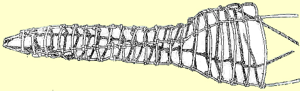
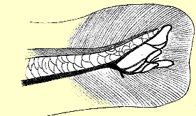
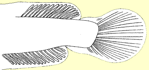
The similar habits of "lula" and the existence of runways to and from the nests are known to some of the natives of this country, who utilize the knowledge for the purpose of capturing the fish during the breeding seasons. They say that whilst watching over its eggs and fry, "lula" will not take the bait and can only be caught by the kuda This is a small basket of deep conical form and wide mesh, about 20 inches long, ending blindly at the narrow end, opening like a funnel at the wide end, just large enough to receive the body of the fish, which, once inside, cannot withdraw. At Minneriya these lula-kudu are made by a cunning old descendant of the Veddas, named Sirataweli, and they are also used at Topawewa. (See figure below.)
| Vedda trap for nesting lula, in use at Minneriya (1/4 of size) |
On May 28 a man brought some "lula" eggs to me, which he had taken from a nest amongst the rushes in the Hunupitiya arm of the Colombo Lake. I waded out to this nest and again saw the characteristic translucent golden yellow or ambercoloured eggs, some newly hatched, spread like a sheet, flush with the surface in a subcircular area behind a tussock of rushes which partly served to filter the direct rays of the sun. Amongst and around the eggs were scattered the usual detached fragments of herbage, consisting chiefly of small leaves of aquatic plants. 1 did not see the adults, but the man said that both parents had been near the nest, the smaller of the two aggressively protecting it; he called this one the female, possibly not knowing that amongst fishes the male is smaller than the female.
At a little distance from the "lula" nest just described there was a swarm of "mada-karaya" fry. A few days later (June 3) another batch of the same kind of eggs was brought to me from Hunupitiya, but tinder said that they were "mada-karaya" eggs, and lie brought a dead fish of this species along with him to prove his point. They had the same diameter, about 1.25 mm., as the "lula" eggs, and the subsequent stages were those of the development of lula." 1 conclude therefore that 1 have not yet seen the spawn of "mada-karaya", and do not know for certain whether the eggs of this species float at the surface like those of "lula", or whether they lie at the bottom like those of most other freshwater fishes. I hope to clear up this point at an early date.
On June I still another lot of "lula" eggs was brought in from Welikada, near the toll-bar oil the road to Kotta near Colombo. The material upon which this paper is based has thus been derived from four broods, namely one from Minneriya - two from Hunupitiya and one from Welikada.
The floating eggs of "lula" owe their buoyancy to the presence of a single large oilglobule which occupies the greater part of the ovum and is immersed in the golden yellow yolk (Fig. 1).
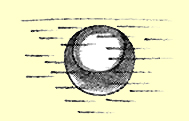 | Fig. 1.- Egg with single large oil-globule, floating below the surface film of water, as seen with a simple lens. |
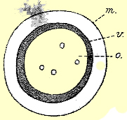 | Fig. 2.- The same seen from the upper pole under a low power. The small spherules inside the large globule appeared with a deep focus. m,. vitelline membrane; v, yolk; o, oil-droplet. |
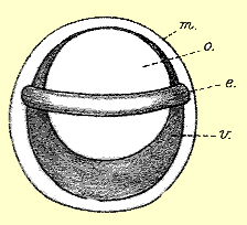 | Fig. 3.- Egg seen in suspension about fifteen hours later; e, embryo. |
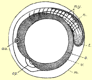 | Fig. 4.- The same seen from above; op, eye; au, auditory sac; my, myotomes; n, notochord; t, free end of tail. |
A few hours later the heart begins to beat and the tail to twitch. Tile orientation of the embryo is constant, the left side being uppermost when viewed from the upper pole of the egg (Figs. 4 and 5). The movement of the tail soon lead to the rupture of the vitelline membrane and the liberation of the embryo; and now commences the larval period of development.
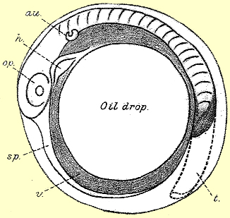 | Fig. 5. - Egg five hours later, from above. The shaded portion of the tail (t) shows it bend down, the dotted portion shows it in extension. h, heart; sp, body-cavity (splanchnocoel); v, yolk; other letters as before. |
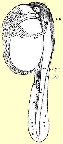 | Fig. 6. - Newly hatched larva; length 3,5 mm.; eyes colorless. pc, posterior cardinal vein joining the caudla vein to form the subintestinal vein; ao, aorta; au, auditory sac containing two otoliths. |
For three days after hatching the larvae remain at the surface of the water, resting on one side with the yolk-sac Lip. When they swim they sway and rotate irregularly, sometimes apparently spinning round. The network of stellate cells over the yolk-sac soon darkens and forms a pigment reticulum, and pigment begins to appear in the eyes on the first day, but, as I have already mentioned, at the time of hatching the eyes are clear and devoid of pigment. In fact, throughout a great part of the first day after hatching, the spawn presents the same appearance as it did before hatching, and only closer inspection reveals that the yolk-laden larvae have escaped from the egg-membranes. First day hatchlings are thus distinguished as unpigmented or clear "lula" surface hatchlings.
Second and third day hatchlings are called pigmented "lula" surface hatchlings. They measure 4-5 mm. in length. Whereas in the very young hatchling the caudal vein passes in toto alongside the hind-gut into the subintestinal system (Fig. 6), it is now seen to give rise to a capillary system which is joined by the posterior cardinal vein and discharges into the subintestinal vein behind and below the yolksac (Fig. 7).
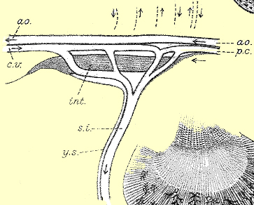 | Fig. 7. - Second day larva. Sketch of the capillary network at the confluence of the caudal, posterior cardinal, and subintestinal veins. The arrows above the aorta indicate the direction of the flow of blood in the segmental parietal vessels. ao, aorta; cv, caudal vein; pc, posterior cardinal vein; int, intestine; ys, posterior surface of yolk-sac ; si, subintestinal vein running round the yolk. |
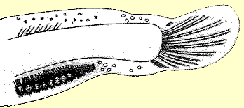
 | Fig. 11. - Twelfth day larva. View of the tail end from the left side, showing notochord bending up and a capillary formation below it, the finely radiate structure of the embryonic fin, and its truncate extremity; nc, medullary tube; nch, notochord; cv, caudal vascular system, diagrammatic, without distinction of artery and vein. |
By the third day the length is slightly increased, about 5 mm., the pigmentation is intensified, and the mouth is open. Besides the uniform layer of pigment over the yolk-sac, stellate cells are distributed over the myotomes, especially along their upper and lower borders, and a thin line of cells occurs along the centre of the dorsal and ventral embryonic fins, which at this stage show an equal amount of pigment. On this day indications of the air-sac appear, the pectoral fins begin to flap, and respiratory movements commence. The larvae still rest at the surface when not swimming, and are incapable of resting at the bottom.
On the fourth day the larvae have attained a length of 6.75 mm. They are now leaving the surface and frcely at all levels; bright yellow spots appear over the eyes. The hinder extremity of the notochord is still straight. but the embryonic fin has exchanged the vacuolated structure of the preceding days for a fine radiate striation. The sides of the body are free from pigment, and are consequently traversed by a pale longitudinal band parallel with the notochord. There is also a more or less interrupted pale band in the middle dorsal line of the fore-body, in front of the embryonic fin. There is a faint massing of embryonic tissue below the hinder end of the notochord, a little before its extremity. This is the primordium of the caudal fin-ray system.
The fifth day shows no increase of length. The caudal pigment is increasing, though still diffuse, and the caudal primordium is becoming denser. On the sixth day the length is found to exceed 7 mm., and there is a further slight concentration of pigment and embryonic tissue.
On the seventh day, still with a length of about 7 mm., we find the first traces of the basal cartilages of the caudal rays, situated below the free straight end of the notochord at the point where the myotomes or muscular segments cease. The rudiments of three cartilages can be made out, but the caudal pigment, which has a peculiar relation to the formation of the fin-rays, is still diffuse. The pigment in the ventral portion of the embryonic fin is now beginning to predominate over that in the dorsal portion of the fin.
On the twelfth day (Fig. 11) the end of the notochord begins to bend up, and the caudal rays begin to show; there is still no further increase in length; on the contrary, as the tissues and cavities of the body begin to expand, there is a very slight decrease in length to be noted, from about 7 mm. to about 6.75 mm., this length being maintained to the end of the fifteenth day. Incidentally I noted the fact that the twelfth day larvae were not rising to the surface of the water to take the air. Up to this time the body of the larva has been colourless, except for the black pigment. On the fifteenth day some of the more advanced larvae display a pronounced yellow ground colour associated with a further condensation of the black cells. The contour of the caudal fin at this, period is shown in Fig. 11.
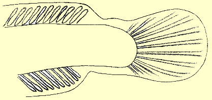
There is now a gap in my observations until the twenty-fifth day, the fry having been kept meanwhile within a small enclosure in a glass tank. The embryonic fins are still continuous (Fig. 12) the ventral portion of the embryonic fin, from which the anal fin arises, is much more highly pigmented than the dorsal portion; neither of them shows any trace of secondary rays.
 | Fig. 12. - Twenty-fifth day larva. Tail end from the left side. The total length of the larva was 7,5 mm. The figure illustrates the commencing fasciculation of the caudal striations, and the basal cartilages below the subterminal convexity of the notochord. df, dorsal embryonic fin; m, muscle fibres below the notochord; vf, ventral embryonic fin; more highly pigmented than the dorsal. |
Larvae thirty-seven to forty days old vary in length from 10 to 13 mm. The length bears no direct proportion to the bulk, since a larva of 13 mm. has at least twice the bulk of one of 10 mm. This is the transition period from the larval to the postlarval phase of growth. At 10 mm. there are still no external rudiments of the ventral fins, but these appear when the larva has attained the length of 10-25 mm. They arise in situ a short distance behind the plane of the pectoral fins and far in front of the vent, as minute buds close to the middle line of the abdominal surface. The embryonic fins are still continuous, but a shallow constriction, both above and below, separates the future definitive dorsal and anal fins from the caudal fin, the intervening portions of the embryonic fin in the region of the caudal peduncle undergoing reduction and degeneration, accompanied by the appearance of vacuoles in their substance (Fig. 14). The primordial formative tissue of the dorsal and anal rays has now invaded the corresponding parts of the embryonic fin, obscuring the striations, leaving a peripheral rim free, where the primary striations are clearly visible.
 | Fig. 14. - Thirty-seventh day larva; length 10 mm. Posterior end of the body to show the commencing constriction of the caudal fin and the primordia of the dorsal and anal rays, the latter accompanied by dense pigment. |
The larvae are now - swimming near the bottom of the shallow aquarium in which they have been reared and come to the surface to take in air. On the twentyeighth day a larva gulped the air once a minute, eight times in eight minutes, each time leaving a small air-bubble, at the surface. As already indicated, the fortieth day after hatching may be taken to mark approximately the end of the larval development in so far as this is denoted by the condition of the tins (Figs. 15 and 16).
 | Fig. 15. - Fortieth day; length 10.75 min. Constriction of caudal fin progressing. |
 | Fig. 16. - Hinder end of another larva of the fortieth day; length 12.5 mm. Constriction of caudal fin complete. |
The subjoined table gives a summary of the chronological data which I have been able to ascertain regarding the external features of the development of Ophiocephalus striatus
| Day of Life | Total Length | Principal Events |
| 1st | 3 to 5 mm. | Yolk-sac circulation established pigment cells develop their black colouration; pigment begins to appear in eyes. |
| 2nd to 3rd | 4.5 to 5 mm. | Pectoral fins arise; mouth opens, and respiratory movements commence |
| 4th | 6.75 mm. | Larvae leaving the surface and swimming freely at all levels. Bright yellow spots over eyes |
| 7th | 7 mm. | Larvae swarming and turning in unison at the slightest concussion. Caudal cartilages appear |
| 12th to 15th | 6.75 mm. | Posterior end of notochord bends up |
| 28th | 8 to 10 mm. | Caudal rays jointed and articulated with the basal cartilages.Larvae rise to surface to take air |
| 37th | 10 mm. | Primordia of dorsal and anal rays |
| 40th | 10.25 to 13 mm. | Rudiments of ventral fins appear Dorsal and anal fins separating from caudal |
| 63rd | 17 mm. | The fry now hide in the mud |
| 73rd | 25 mm. | The fry now hide in the mud |
With reference to the above it is to be noted that after the absorption of the yolk about the fifth day after hatching, when the larvae begin to feed independently, the daily growth begins to vary; and the variation is probably the greater on account of the larvae having been reared in captivity. It has been similarly noticed that in the case of the trout, larvae of equal ages occur in very different stages of development. 1 Only one larva was examined on the thirtyseventh day. The striking increase in length to the extent of a millimetre between the first and second days is normal and noteworthy.
On July 28 a swarm of "lula" fry was found close to the bank in a part of the Colpetty arm of the Colombo Lake. They were incessantly streaming to and from the surface, and their presence in the thick water was made manifest by this action. They were coloured a soft reddish brown or brown and pink, quite different from the black and yellow of "madaya" fry (O. punctatus) of the same size. Their total length (including the caudal fin varied from about 15 to about 1.7 mm. - a size which corresponds with the dimensions of larvae which I have reared in an aquarium for sixty-three days after hatching. The general colour-effect is dominated by abroad lateral reddish orange band occupying almost the entire height of the myotomes, commencing from the eye on each side, and ending behind with a rounded edge at the base of the caudal fin concentric with the terminal contour of the latter. The iris is golden with a red flush; there is a bright golden occipital point; and the basis of the anal fin is dense black along its whole length. The colour of the fry is essentially that which it had acquired at half the size; and it retains this colour until it has doubled the size, after which the definitive markings begin to appear. 2
On the same occasion (July 28) I obtained a sample of "madaya" fry of the same general dimensions as the "lula" fry from a neighbouring muddy swamp. Instead of the reddish brown subtranslucent ground colour of the "lula" fry, the "madaya" fry are characterized by a blackish ground colour, upon which the bright golden yellow bands stand out clear, namely, a pair of lateral bands about half the width of the "lula" bands, occupying the central third of the height of the myotomes and ending behind in a point extending about one-third of the length of the caudal fin into the substance of the fin. Along the length of the back is a golden yellow line running along the basis of the dorsal fin, and presenting a more or less distinct interruption in the occipital region in front of the fin at the spot where there is a minute golden speculum in "lula". Besides all this, the- madaya fry present a clear yellow spot on the snout and do not possess the black basis of the anal fin. The postlarval stage of "lula" is thus easily distinguishable from the postlarval "madaya". Moreover, at this stage the median fins have completed their differentiation from the embryonic matrix. - "lula" and 11 "madaya" fry of 17 mm. exhibit the following differences in respect of their median fin-rays and lateral bands
| name of fish | number of dorsal rays | number of anal rays | lateral band with |
| "Lula" | 47 | 30 | 1.2 mm. |
| "Madaya" | 30 | 20 | 0.6 mm. |
So far as I am aware no other instances of eggs of freshwater fishes floating at the surface of the water by their own buoyancy have been described hitherto. The same advantages. namely, direct proximity to atmospheric air and to sunlight, are partly secured in other ways, as by attachment to aquatic plants or by deposition in very shallow water. The floating eggs of Ophiocephalus striatus, with their accompaniment of small fra-7nients detached from the surrounding plants, such as cut pieces of tank weed, leaves, and lengths of cut stem, rather invite comparison with the floating nests of Gymnarchus which were described by the late J. S. Budgett.3 He found these nests amidst the dense grasses of a West African swamp floating at the surface in three to four feet of water; the deepest part of a nest was only about six inches below the surface; in it "were deposited about a thousand large spherical amber-Eke eggs 10 mm. in diameter. The eggs hatched five days after being laid, and in eighteen days a thousand young fry of Gymnarchus niloticus left the nest" having in this short time attained a length of three inches.
In the same flooded grass-lands Budgett frequently found "masses of white foam floating on the surface of the water." These proved to be the foam-nests of another fish, Sarcodaces, a member of a family, the Characinidae, which is not represented in Ceylon. They were filled with numerous transparent ova, 2.5 mm. in diameter; on 2 hatching, the larvae "make their way through the foam in which they are laid, down to the surface of the water, and there the young larvae hang, holding to the surface of the water by a large adhesive organ situated on the front of the head." There is no adhesive organ in the young larval of Ophiocephalus striatus, but all the same they are kept at the surface by the extraordinary buoyancy of the yolksac ' and in due time they strike out from the surface to the bottom; whereas the hatchlings of most freshwater fishes strike out from the bottom towards the surface.
Thus the larva of Ophiocephalus differs from that of Sarcodaces in the absence of a frontal cement organ, and from that of Gymnarchus in the absence of external gills. Like the Mormyridae, the family to which Gymnarchus belongs, and the Characinidae, the Ophiocephalidae is a characteristic family of tropical freshwater fishes whose morphology is better known than their bionomics; and the present contribution, with others which are to follow, will show how much remains to be done upon the subject which was so successfully and tragically inaugurated by Budgett.
There are several other points with regard to which comparisons with other forms may be both instructive and interesting. The simple ventral flexure of the embryo of Ophiocephalus, the absence of retinal pigment within the egg, and the formation of the pectoral fins after hatching, are facts to be noted as contrasting with what, occurs in the development of many other Teleostean fishes. The later appearance of the ventral fins is the rule amongst bony fishes.4 In the two Dipnoan fishes, Lepidosiren and Protopterus, the paired limbs arise simultaneously; in the former thirteen days after hatching; 5 in the latter, according to Budgett, the rudiments of the limbs begin to show about the third day after hatching, and by the tenth day after hatching the larva is provided with well-developed limbs. 6 On the other hand, in the Australian Dipnoan genus Ceratodus, according to Semon's observations, 7 the ventral fins appear about a month later than the fore-limbs, approximating in this respect to what I have described above for Ophiocephalus. In this connection it is interesting to note that in the genus Channa which also belongs to the Ophiocephalidae and occurs in Ceylon, the ventral fins fail to put in an appearance throughout life.
As described above, the development of the definitive caudal rays of Ophiocephalus is marked by a succession of phases: firstly, the proliferation of primordial or formative tissue below the posterior end of the notochord; then the formation of a capillary plexus; the development of basal cartilages and the fasciculation of the primary striations; lastly, the appearance of the basal prongs. The actual rays thus arise peripherally or centripetally; and this relation appears to be more pronounced in the case of the anal rays (Fig. 14). An analogous peripheral origin of fin-rays has been described by J. Schmidt in the pelagic larvae of the marine salmonoid fish, Argentina silus, where at a stage of 28-32 mm. in length, the primordia of the dorsal and anal interspinous rays appear nearly halfway between the contour of the body and the outer border of the embryonic fin, without direct connection with the body. At a length of 39 mm. the fin-rudiments touch the margin of the body. 8 The general ontogeny of the median fins of Ophiocephalus resembles that described and figured by Assheton for Heterotis niloticus. 9
The rhythmical darts to the surface for the gulping of air by the 28-day larvae of Ophiocephalus is paralleled by the larvae of Gymnarchus, although the latter are burdened by an enormous pendent or external yolk-sac which persists much longer than the yolk-sac of Ophiocephalus. "By the tenth day after hatching the larvae are able to drag their yolk-sac to the surface of the water, when they take a gulp of air into their lunglike swim-bladder and fall again to the bottom, on reaching which they again start for the surface with unceasing regularity, so that when looked at from above, the nest of Gymnarchus, with its swarm of scarlet-bearded [referring to the external gills], yolk-hampered larvae, presents a most amazing spectacle." (Budgett, op. cit. pp. 131 - 32.)
The circulation of the blood in the larvae of Ophiocephalus conforms to the larval Teleostean type of circulation and presents several interesting characteristics, chief among which are the direct junction, during the early days of larval life, between the aorta and the caudal vein; the yolk-sac circulation which is intercalated in the subintestinal system in a manner analogous to the relations of the hepatic coecum in Amphioxus; the subintestinal vein itself, which, as was pointed out long ago by Balfour (op. cit. p. 651), recapitulates an ancient pre-piscine organization; lastly, the reversed current of the posterior cardinal blood, which, instead of flowing forward towards the heart, flows backwards to join the subintestinal system, so that the aortic blood and the cardinal blood is seen to be flowing in the same direction.
Lastly, it may be noted that although, as I believe, the floating eggs of "lula" are unique amongst freshwater fishes, so far as the available records go, yet they present some analogy with the egg's of some species of marine fishes. Thus the transparent eggs of the greater weever (Trachinus draco), with a diameter varying from a little less to something over one millimetre, have a single large oilglobule and float at the surface of the sea. Four or five days after oviposition the embryos are hatched; and four or five days after hatching the yolk has become absorbed. The buoyancy of the yolksac causes the larvae to float helplessly in the water for some time after hatching, with the yolk-sac uppermost. 10
The usefulness of "lula" as a source of food-supply for the lowcountry of Ceylon, together with the fact that attention is now being drawn to the maintenance and replenishment of the stock of fishes in the rivers and tanks of this country, makes it incumbent upon us to examine the practical bearing of the foregoing observations and others like them. In other places it is recognized that methods of culture should be based upon a knowledge of the breeding habits of fishes under natural conditions,11 and if this point of view is accepted locally the utility of these notes on "lula" may be taken for granted. The necessity of differentiating between the successive ages and stages of the growing fish, and between fry of the same age belonging to species which may be closely allied zoologically though far apart economically; and the study of the conditions under which fry can be reared best under an artificial system, are points which must always guide cultural operations whenever they are undertaken.
The feeding of "lula" during its earlier stages is not an insuperable difficulty, and under suitable conditions it even goes forward to a large extent automatically. It is known that the growth of fishes is governed directly by the food-supply. "Lula" is one of those fishes whose size-limit is practically indefinite. The more food it receives of the right kind, the quicker and the larger it grows. Some young "lula" which I kept in an aquarium at my bungalow in Colombo had an average total length of about 35 mm. in February, 1908; 45 mm. in July, 1908; 96 mm. in April, 1909. The series last measured consisted of six individuals ranging from 85 mm. to 115 mm. The latter measurement may be taken as representing approximately the usual growth of a yearling "lula", although under more favourable conditions it might reach six inches in total length front the tip of the snout to the end of the tail fin. It will be observed that in the nine months from July to April the young "lula" more than doubled their length. A sample of "kavaya" fry, Anabas scandens, six in number, kept in the same tank at the same time, behaved in a similar manner; when first measured in June, 1908, their average length was 33 - 5 mm.; a month later the average had increased to 41 mm.; and in April, 1909, it had reached 83 mm. Another tropical freshwater food-fish, which may be introduced into Ceylon some day, namely, the Gourami (Osphromenus olfax), is known to attain a length of about four inches in the first year, seven or eight in the second, and ten or eleven in the third, after which it begins to breed. 12
From what has been said above, and also from what has long been known respecting the powers of endurance possessed by the Ophiocephalidae, it is obvious that, as soon as required, yearling "lula" could be reared with comparative ease in protected ponds, and could be distributed subsequently as required.
The extent to which the fry of freshwater fishes depend for their sustenance upon the aquatic larvae of mosquitoes or Culicidae is a matter of practical moment and also of special interest at the present time, for although the little fishes called" millions" in the West Indies, topminnows elsewhere, are not such a universal panacea for malaria as it has been suggested they would prove to be, yet it is useful that renewed attention should be given to the part played by fishes in general, by the fry of the larger fishes, and by top-minnows in particular, in checking the natural increase of mosquitoes by feeding upon their larvae.
Mosquito larvae too often thrive best in places to which fishes can never have access, as in the small accumulations of chocolatecoloured water in the crevices of jungle trees; and even in the tanks they favour the scum-covered water at the edge, where the vegetation is rotting, though this of course will vary with the waterlevel in the tank at a given time. An analogous case is afforded bythe Colombo Lake fly, an undetermined species of Chironomus, whose numbers have not appreciably diminished in spite of all the precautionary steps, which have been taken during the past five or six years, due, it must be supposed, to the fact that although their larvae are naturally useful as fish-fodder, yet they thrive best in places which are not otherwise attractive to insectivorous fishes. In such a case as this the normal balance cannot be restored without the application of herculean methods.
Next to the microscopic crustacea which compose the most important source of food-supply for the young of freshwater fishes, the larvae of mosquitoes are looked upon as the best friends of the fish-culturist, although the mature insects are amongst our worst enemies. 13 We are thus placed, often at one and the same time in perplexity as to flow we may procure the larvae and how to avoid the flies. In any case, it is expedient to recognize as beneficial those species of fishes which prey upon mosquito larvae.
Wishing to test the selectivity of mosquitoes for different waters, I placed two chatties side by side, one (referred to m A) containing water rendered turbid by decaying animal matter, the other (B) containing clear water. Two days later 1 took 20 culicine egg-rafts from A, as against 1 from B. On the following day 11 more eggrafts appeared on A; none on B. On the day after this, again 14 rafts were found on A, only 2 on B, the water not having been changed in the meantime. At the same time another species was observed to lay single eggs which adhered to the surface of chatty A below the surface of the water close to the water's edge. Water containing decaying vegetable matter is equally attractive to eggladen mosquitoes. Such a pronounced preference for a putrescent nidus for the eggs on the part of insects whose feeding habits are so highly specialized, amounts to a biochemical reaction to which the term saprotaxis might he applied.
Young Anabas not only consume the larvae, but will swallow the entire egg-rafts, each comprising about 160 eggs. On June 4, "lula" fry of the seventh day after hatching were observed to ingest a quantity of separate white mosquito eggs floating on the surface of the water in a glass dish. Here it may be mentioned that on the same day about 30 "lula" surface hatchlings from another brood were eaten up by older "madaya" fry. Later on it was found that the "lula" fry would approach the culicine rafts, but would not eat them; as soon however as the minute larvae hatched out from the eggs, the fry devoured them. Since then I have fed my "lula" fry with abundant mosquito larvae, without always observing the actual process of ingestion.
Top-minnows (Cyprinodontidae) are represented in Ceylon by one or two species of Haplochilus, of which the commonest is H. lineatus called "diya pita hendeva" in Sinhalese. They lay eggs which become attached by glutinous threads to water plants, about as large as "lula" eggs, with pale amber-coloured yolk containing a number of oil-globules. I have not found them so attached, but have seen them freshly extruded in July. The vitelline membrane shows a reticulated sculpturing, and the long adhesive threads radiate from a centre placed near the oilpole. The mature fish attains a length of about I I inch; the snout is flattened and shovel-shaped, adapted for surface feeding; the male is larger than the female, exhibiting at the breeding season a bright golden green lustre on the so ales of the hind-body; ventral and anal fins greenish, the latter with backwardly prolonged orange-coloured rays and three black basal flecks : dorsal and caudal fins and lower lip orange; hind-body without the 6-8 vertical black bars which are present on the spawning female. Maimed mosquitoes dropped upon the surface of the water are seized and swallowed by the top-minnows. These fishes (H. lineatus) possess a flashing white occipital triangular spot, which can be alternately darkened and rendered invisible by the expansion of black pigment cells, and can again flash out resplendently when these contract. It is not phosphorescent; I think that it acts as a lure, but have not been able to prove that it does.
This article originally appeared in: Spolia Zeylanica, vol. VI, Part XXI, March 1909, pp. 108-123. Colombo, Sri Lanka.
1Compare E. Goeppert. Die Entwicklung des Pankreas der Teleostier Morph. Jahrb., XXI., 1893, p. 90. Back
2 A. Willey. Fishery observations. Spol. Zeyl., V., 1908, p. 145 et seg. I take this opportunity of correcting the name of a protozoon referred to in this note as Blepharisma. It should have been Loxodes Back
3 J. S. Budgett. On the breeding habits of some West African fishes. Trans. Zool. Soc. London, XVI.. 1901. Reprinted in the Budgett 'Memorial Volume edited by J. Graham Kerr. Cambridge Univ. Press. 1907, pp. 119-36, plates and text-figures. Back
4 F. M. Balfour. Comparative Embryology, 2nd edit., 1885, Vol. 11., p. 80. Back
5 J. G. Kerr. Development of Lepidosiren paradoxa Phil. Trans. R. Sec., London, Vol. 192, 1900, p. 316. Back
6 J. S. Budgett. Op. cit. Pl. VIII., figs. 12-13, p. 127. Back
7 R. Semon. Forschungsreise. quoted from Kerr, toe. cit. Back
8 J. Schmidt. Development of the Arjentines. Medd. fra Komm Fiskeri 11. , No. 4, Copenhagen, 1906. Back
9 R. Assheton. Teleostean Larvae from the Gambia River. Budgett Memorial Vol., Cambridge, 1907, p. 439. Back
10 J. Boeke. On the early development of the Weever Fishes. Tijdschr. Nederland. Dierk. Ver (2) VIII., 1903, pp. 148-57, Pl. VII. I have not seen the original paper, but it is quoted by Dr. Theodore Gill in "Life histories of Toadfishes compared with those of Weevers and Stargazers". Smithsonian Miscellaneous Collections, Quarterly Issue, Vol. III.. Part 4, Washington, 1907, p. 421. A subsequent paper by Boeke on "The later larvael development of Trachinidae" appeared in the above-named journal. Vol. X. 1907, pp. 245-54. Back
11 Compare Dwight Lydell. The habits and culture of the Black Bass. U. S. Fish Comm. Bull., 1902 (Washington, 1903), pp. 39-44, Pl. S. Back
12 See A. Willey. Ceylon Admin. Rep., 1908. Marine Biology (including first part of Report on Inland Fisheries). Back
13 Compare Dr. Emil Walter. Die Fischerei als Nebenbetrieb des Landwirtes und Forstmannes. Neudamm. 1903, see, pp. 57 62. Back
© 2001 snakeheads.org